
Essential details
MOQ:1
Shipping:Express delivery
Specification Number:FLO-X1
Product Introduction
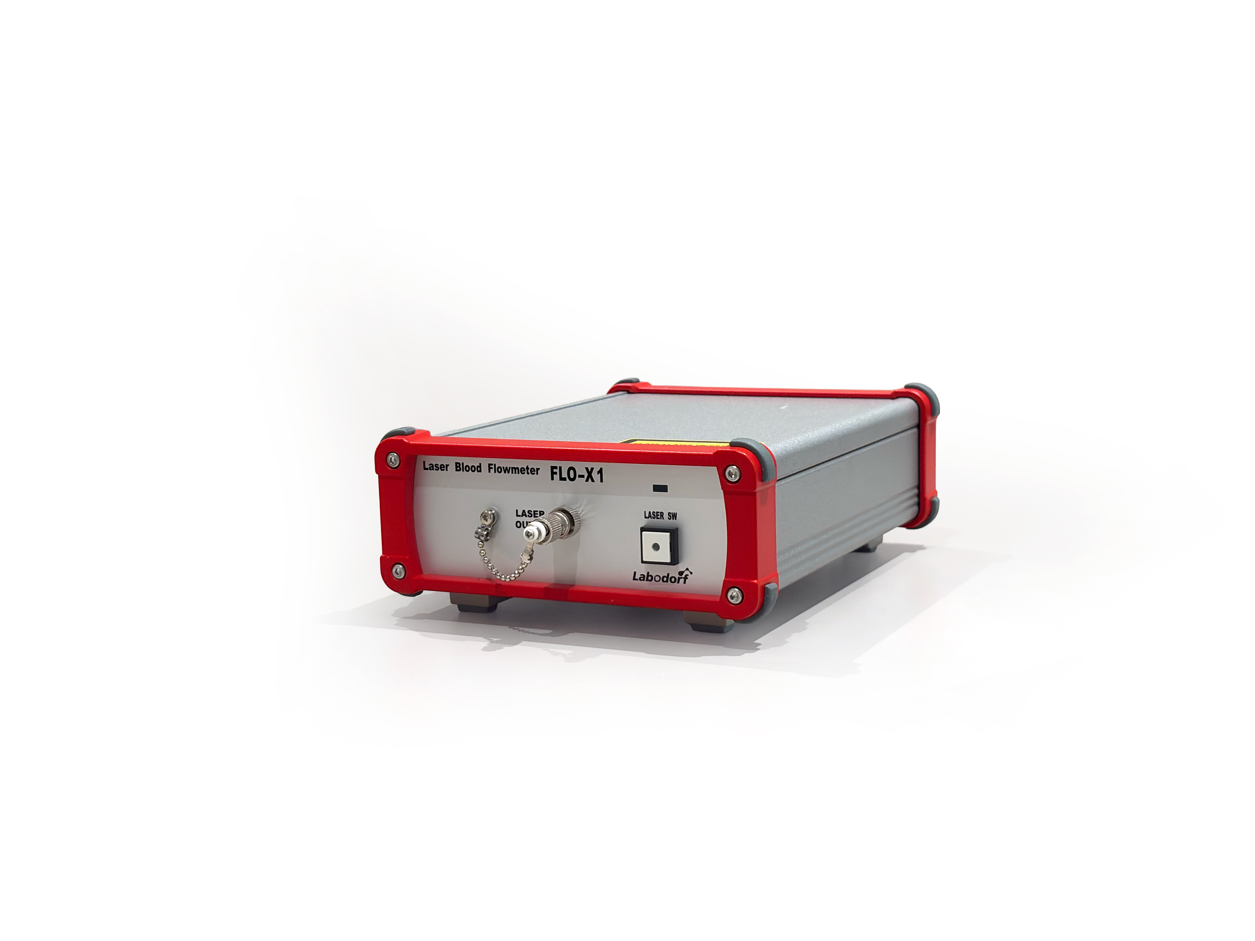
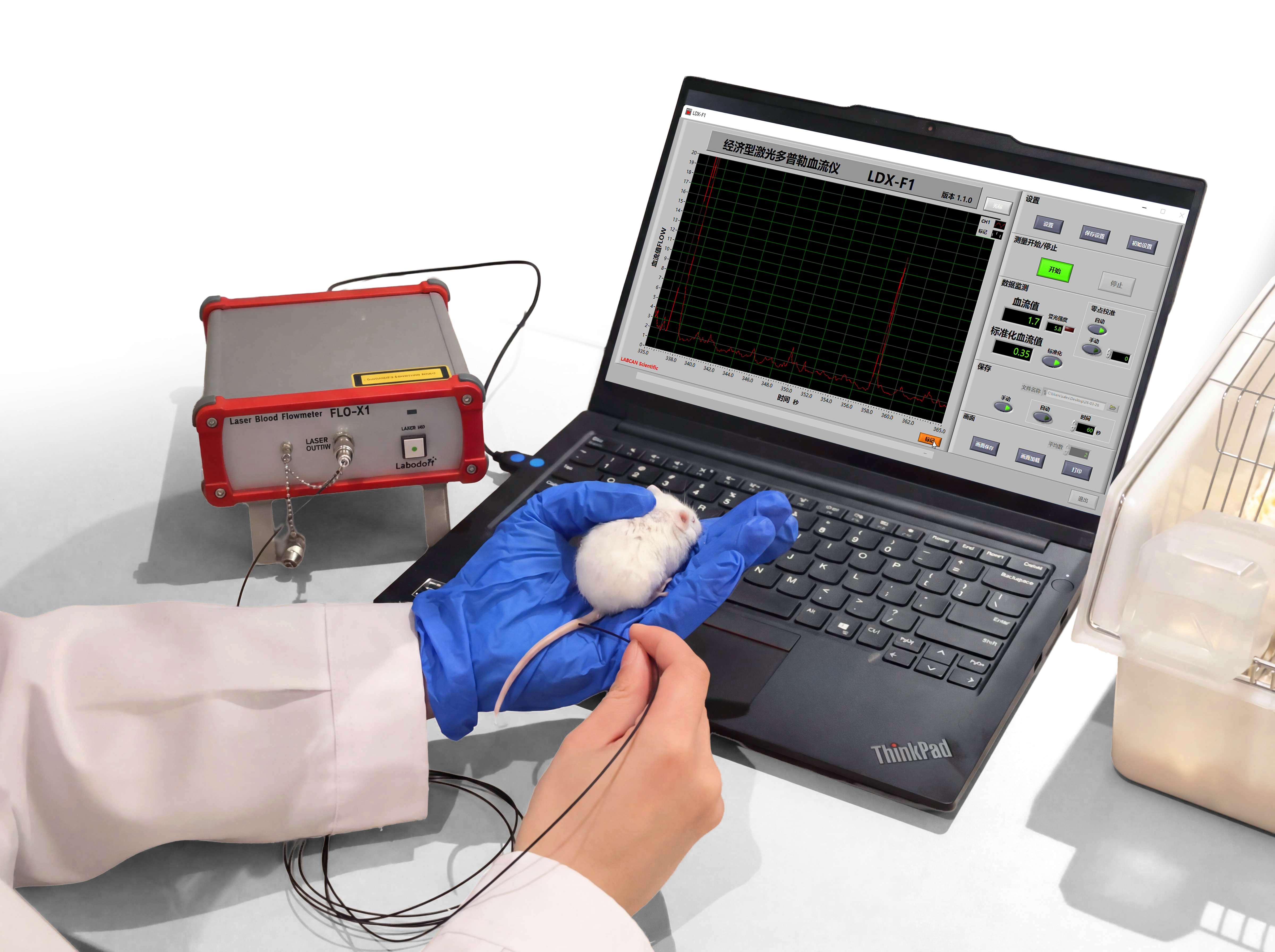
The labodorf FLO‑X1 Conventional Laser Doppler Blood Flowmeter – Mini model is a high‑precision, portable laser Doppler blood flow monitoring device designed for continuous measurement of microcirculatory blood flow in the brain, skin, and other tissues of animals. The system uses a semiconductor laser (wavelength: 780 nm) whose spectral characteristics minimize the absorption difference between oxyhemoglobin and deoxyhemoglobin, ensuring accurate and stable measurements.
Equipped with a standard optical probe, the FLO‑X1 detects microcirculatory blood flow changes at a depth of approximately 1 mm below the tissue surface. The probe uses plastic optical fibers with an ST‑type optical connector for easy connection and flexible operation. The device features a built‑in Fast Fourier Transform (FFT) analysis function, which processes photoelectric signals in real time and optimizes the detection frequency range for more precise and reliable blood flow data. For both basic research and auxiliary clinical applications, the FLO‑X1 delivers a stable and efficient solution for microcirculatory blood flow monitoring.
Working Principle
Light is emitted through the fiber optic probe onto the target tissue, where it scatters widely and is partially absorbed. Some laser light strikes moving blood cells and is reflected back with a shifted wavelength (Doppler shift effect), while light scattered by stationary tissue reflects at an unchanged wavelength. These signals are collected by the receiving fiber, converted into electrical signals, filtered, amplified, and then digitized by an analog‑to‑digital converter to display relative perfusion data.
Features
- Multi‑parameter measurement: tissue perfusion, blood volume, and blood flow velocity
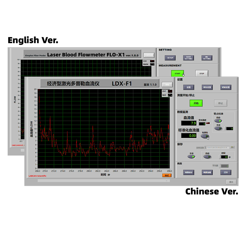
- Continuous blood flow measurement with real‑time display for IMCAO modeling
- Wide selection of probe types for flexible use with different measurement methods and subjects
- Compact size, suitable for various laboratory environments
- Percentage difference in blood flow before and after cerebral ischemic perfusion
Item Specification Model FLO‑X1 Measurement Mode Contact Measurement Area Circular area, approx. 1 mm diameter Measurement Depth 0.5 mm to 1 mm (depending on biological tissue) Tissue Blood Flow 0–1000 ml/min/100 g Detection Frequency Range 0 Hz–50 kHz Time Constant 0.1 / 1.0 / 3.0 s Signal Processor Laptop computer Power Supply 100–240 VAC, 50/60 Hz, approx. 45 W Weight Approx. 1.5–2.0 kg Operating System Windows 10 or Windows 11 CPU AMD 3020e or equivalent RAM 4 GB or more HDD/SSD 128 GB or larger Display Size 11–14 inches Resolution 1366×768 to 1920×1080 Laser Unit Measurement laser: 780 nm, 2 mW, Class 1 (safety level) Laser Connector ST type Optical Fiber 500 μm diameter, plastic silica fiber Fiber Optic Probe Interface: USB 2.0 A/D Resolution 12 bits Probe Power 5 V DC, approx. 0.3 A Dimensions (W×H×D) Approx. 145×60×195 mm Weight Approx. 0.8 kg Operating Environment Temperature: 10–35 °CRelative humidity: 30–90% (non‑condensing)Atmospheric pressure: 800–1060 hPa